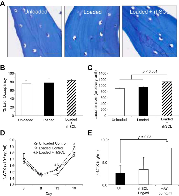
Fig. 6.
Effects of mechanical stimulation and rhSCL on lacunar occupancy and osteocytic osteolysis. Bone cores were processed for analysis after an 18-day experimental period. A: representative toluidine blue-stained sections from each group of bone cores (20× objective; scale bars represent 100 µm). B: the occupancy of osteocyte lacunae was measured from toluidine blue-stained sections; data shown are means ± SE. C: the osteocyte lacunar size at the end of the experiment was measured from toluidine blue-stained sections imaged using a Quantimet imaging system analyzed using ImageJ software. Data shown are means (5 independent sections; at least 100 lacunae/section) ± SE. D: β-CTX levels were measured in culture eluates by commercial ELISA, with each time point representing the concentration of β-CTX in 7 ml of medium after 24 h of perfusion. Data shown are means ± SE. Significant difference: abetween unloaded and loaded controls; bbetween loaded control and loaded + rhSCL groups (P < 0.05). E: β-CTX levels in culture supernatant of human primary osteocyte-like cultures treated or untreated with rhSCL for 72 h. Data represent means ± SD of triplicate wells.

