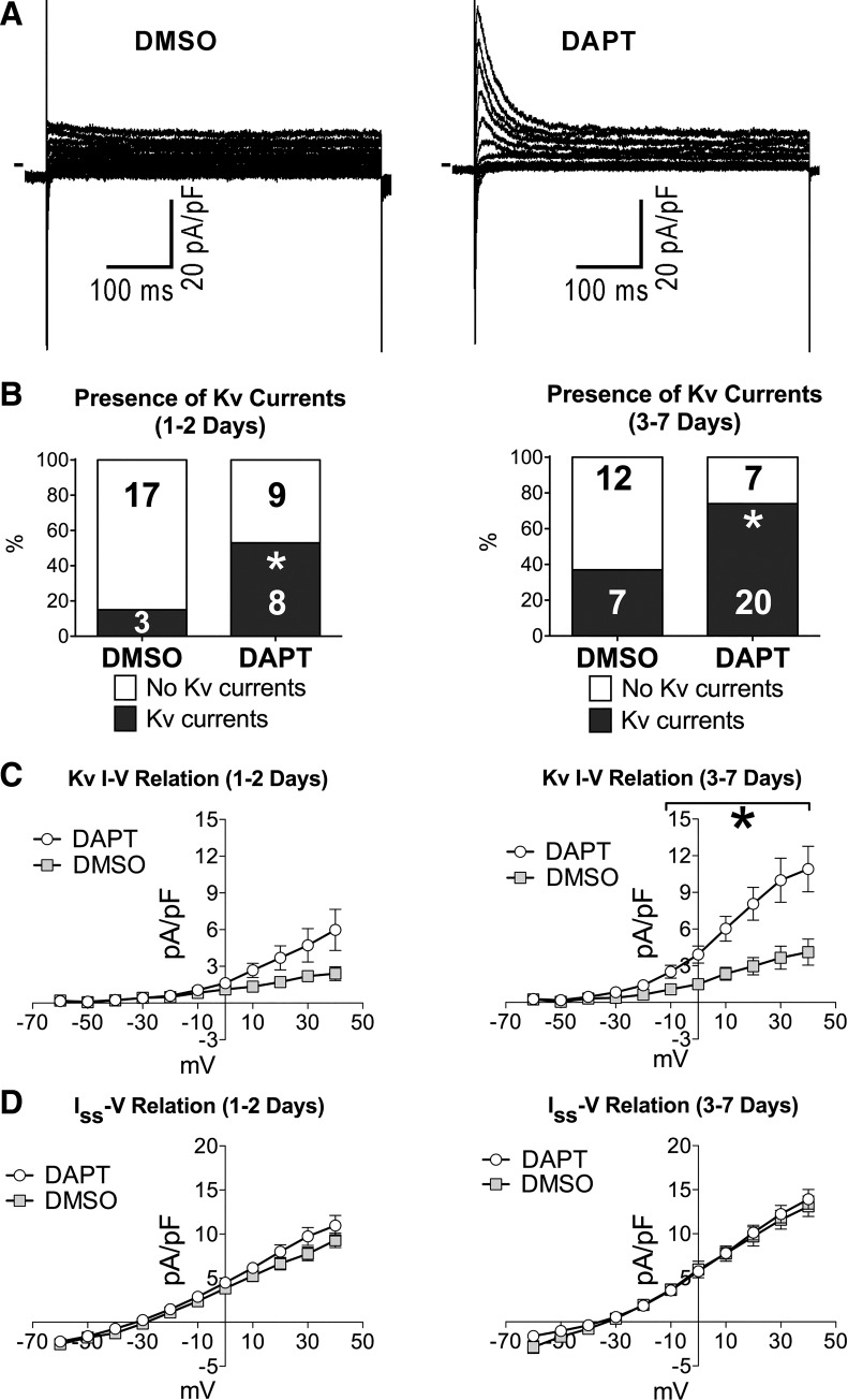
Fig. 8.
Inhibition of Notch1 intracellular domain (NICD) formation in neonatal myocytes promotes Kv currents. A: whole cell voltage-gated K+ currents recorded in voltage clamp in neonatal myocytes in cultures treated with vehicle alone (DMSO) or a γ-secretase inhibitor (DAPT). Currents were elicited by a family of depolarizing steps from a holding potential (Vh) of −70 mV. B: fraction of myocytes presenting outward K+ Kv components in the groups of cells treated with vehicle (DMSO, n = 20 at 1–2 days; n = 19 at 3–7 days) or γ-secretase inhibitor (DAPT, n = 17 at 1–2 days; n = 27 at 3–7 days). The numbers of cells expressing or negative for Kv currents are shown in the graphs. *P < 0.05 vs. DMSO. C: quantitative data for the rapidly activating outward Kv currents in cells reported in B. *P < 0.05 vs. DMSO. D: quantitative data for the sustained current (Iss) measured during the last 50 ms of depolarizing steps in cells shown in B and C.

