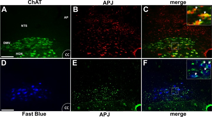
Fig. 1.
Apelin receptors (APJ) are present in the dorsal vagal complex. Representative micrographs showing colocalization of APJ receptors immunoreactive (red) with choline acetyltransferase (ChAT; green; A–C) or neuronal retrograde tracer Fast Blue (D–F; blue) in the intermediate dorsal vagal complex. Insets in C and F are enlargements of the boxes in their respective panels. Arrows indicate colocalized cells in dorsal motor nucleus of the vagus (DMV). AP, area postrema; CC, central canal; HGN, hypoglossal nucleus; NTS, nucleus tractus solitarius. Magnification ×40, scale bars represent 200 μm.

