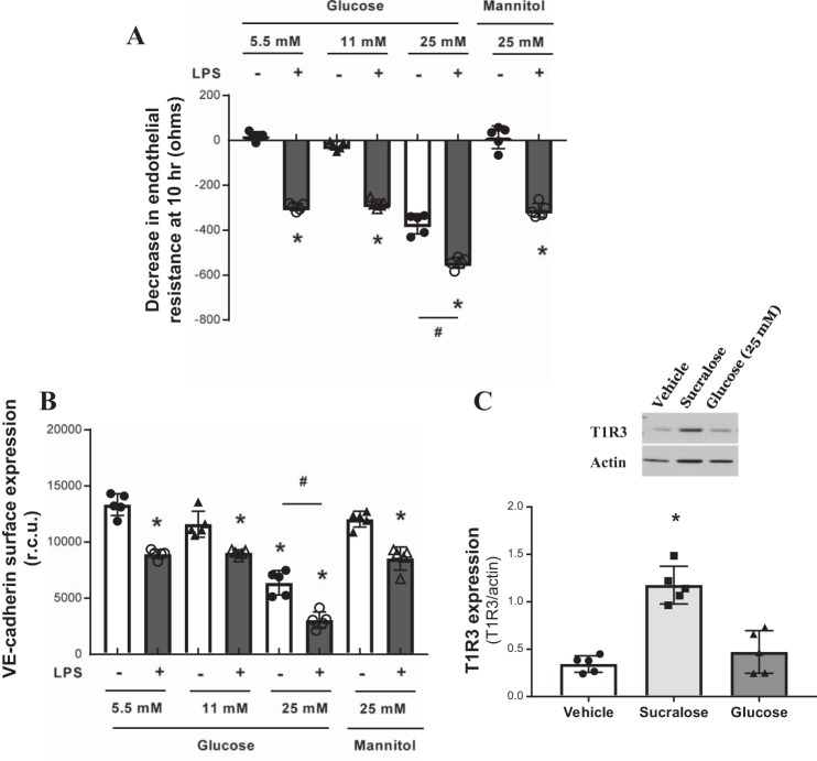
Fig. 4.
High-glucose exposure increases endothelial barrier permeability and VE-cadherin internalisation. A: changes in rat LMVEC endothelial monolayer resistance was measured using ECIS in the presence (closed bars) and absence (open bars) of LPS (1 µg/ml). Monolayers were exposed to different concentrations of glucose (5.5, 11, and 25 mM) or osmotic control mannose (25 mM) at the same time as LPS. Permeability is shown as drop in endothelial resistance measured at 10 h. n = 5. B: cell surface expression of VE-cadherin was determined, with whole cell indirect ELISA using chemiluminescence, following exposure to LPS and glucose as per A. C: protein expression of T1R3 in cultured rat LMVEC exposed to sucralose (0.1 mM), glucose (25 mM), or vehicle for both (H2O) for 24 h. A representative blot (top) and densitometry relative to the load control β-actin (bottom) are shown; n = 5. Data are expressed as means ± SD. *P < 0.05 vs. vehicle for LPS; #P < 0.05 vs. 5.5 mM control.

