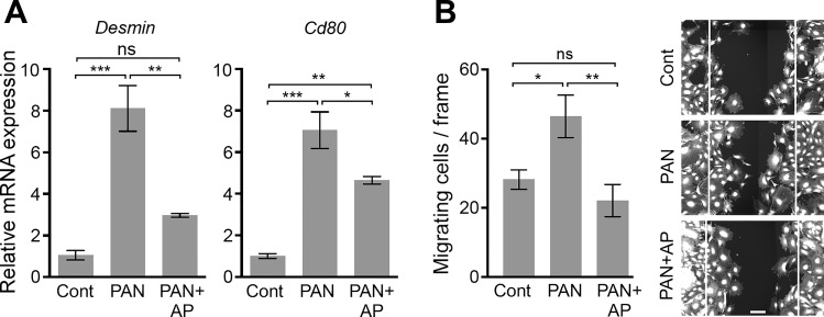
Fig. 4.
Induction of podocyte damage markers is suppressed by alsterpaullone. A: mRNA expression of two known markers of podocyte damage is reduced by alsterpaullone. Graphs showing qRT-PCR-based measurement of Desmin and Cd80 in cultured mouse podocytes treated with either PAN (30 µg/ml) alone or with 30 µg/ml PAN and 1 µM alsterpaullone (AP). Data are expressed as fold change of mRNA levels in treated cells over the control vehicle-treated cells (normalized to 1). Data are means ± SE (n = 3). B: alsterpaullone reduces podocyte migration in scratch wound-healing migration assay. A bar graph showing the number of podocytes migrating toward the gap of scratch in a wound-healing assay (left). Scratch wounds were created in podocyte monolayers by using a sterile 10-μl pipette tip, and the monolayers were incubated in the absence (control, cont) or presence of PAN (30 µg/ml, PAN) at 37°C for 24 h. One set of wounded monolayers was cotreated with 1 µM alsterpaullone (PAN+AP). Subsequently, cells were fixed and stained with CellMask Blue, and the number of cells migrating inside the edge of the wounds was quantified for each condition. Representative images show cells after 24-h treatment (right). The white lines shown in the images represent the wound edges at the beginning of treatment. Scale bar represents 100 μm. The data presented in the graph are plotted as means ± SE (n ≥ 7). One-way ANOVA with Tukey’s multiple comparison test. *P < 0.05; **P < 0.01; ***P < 0.005; ns = not statistically significant.

