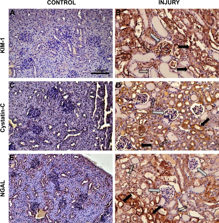
Fig. 2.
Immunohistochemical analysis of cortical tissue in control mice and mice subjected ischemia reperfusion injury of the kidney. A; control cortical tissue probed with kidney injury molecule 1 (KIM-1). B: IRI cortical tissue probed with KIM-1. C: control cortical tissue probed with cystatin C. D: IRI cortical tissue probed with cystatin C. E: control cortical tissue probed with neutrophil gelatinase-associated lipocalin (NGAL). F: IRI cortical tissue probed with NGAL. In IRI images, proximal tubules (solid arrows), distal tubules (open arrows), and glomeruli (hatched arrows) are indicated.

