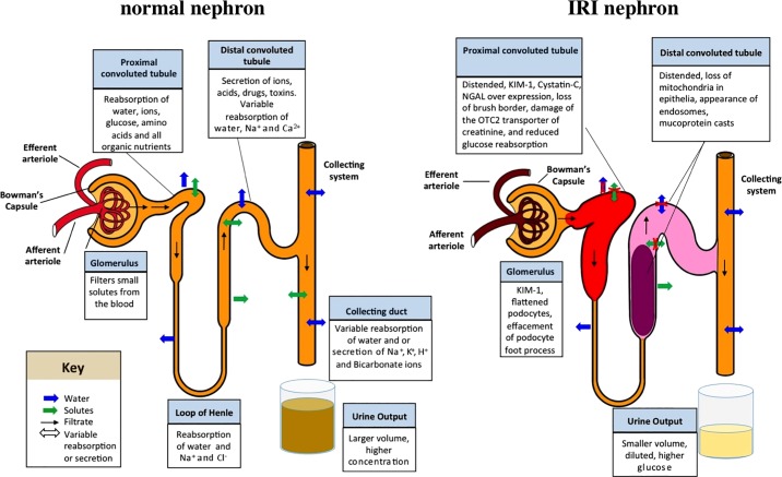
Fig. 7.
Schematic model of a normal and IRI nephron. A: selected normal functions of the nephron are indicated. B: summary of changes to the nephron observed in this study. The red coloration of the glomerulus indicates elevated cystatin C; the widened proximal and distal tubules represent the distension observed in histology analyses; the red color of the proximal tubule indicates elevated KIM-1, cystatin C, and NGAL; the pink coloration in the distal tubule indicates presence of mucoprotein plugs; and the reduced and lighter color of urine in the collection vial indicates reduced urine output and dilution of the collected urine.

