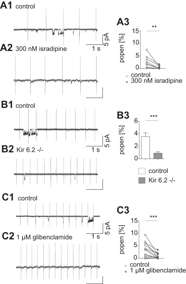
Fig. 2.

Single-ion channel activity is decreased after L-type calcium channel inhibition with 300 nM isradipine. A1: representative current trace before bath application of isradipine; short openings and clusters of channel activity can be detected. The current due to action potentials during pacemaking is clearly visible at regular intervals of several hundred milliseconds. A2: representative current trace during 300 nM isradipine bath application; channel openings appear rarely. A3: mean values for single-channel activity are reduced after isradipine application (Popen control: 2.8 ± 0.85%; Popen isradipine: 0.5 ± 0.24%, P = 0.0078; Wilcoxon signed-rank test; n = 8). B1: representative trace of an on-cell recording from a control animal. Openings of single channels appear in clusters. B2: current trace recorded in a Kir6.2−/− animal. Smaller event clusters can be detected. B3: average Popen recorded in control animals is significantly higher than in animals lacking functional K-ATP channels (Popen control: 3.6 ± 0.58%; Popen Kir6.2−/−: 0.9 ± 0.22%; P = 0.0003; Mann-Whitney test; n = 17). C1: control. C2: representative current trace in presence of the SUR1 subunit inhibitor glibenclamide. C3: glibenclamide causes a substantial drop in channel activation (Popen control: 3.5 ± 0.84%; Popen glibenclamide: 0.9 ± 0.32%; P = 0.0005; Wilcoxon-signed rank test; n = 12). The small 50-Hz oscillation in the current traces is an artifact of the ambient power. **P < 0.01; ***P < 0.001.
