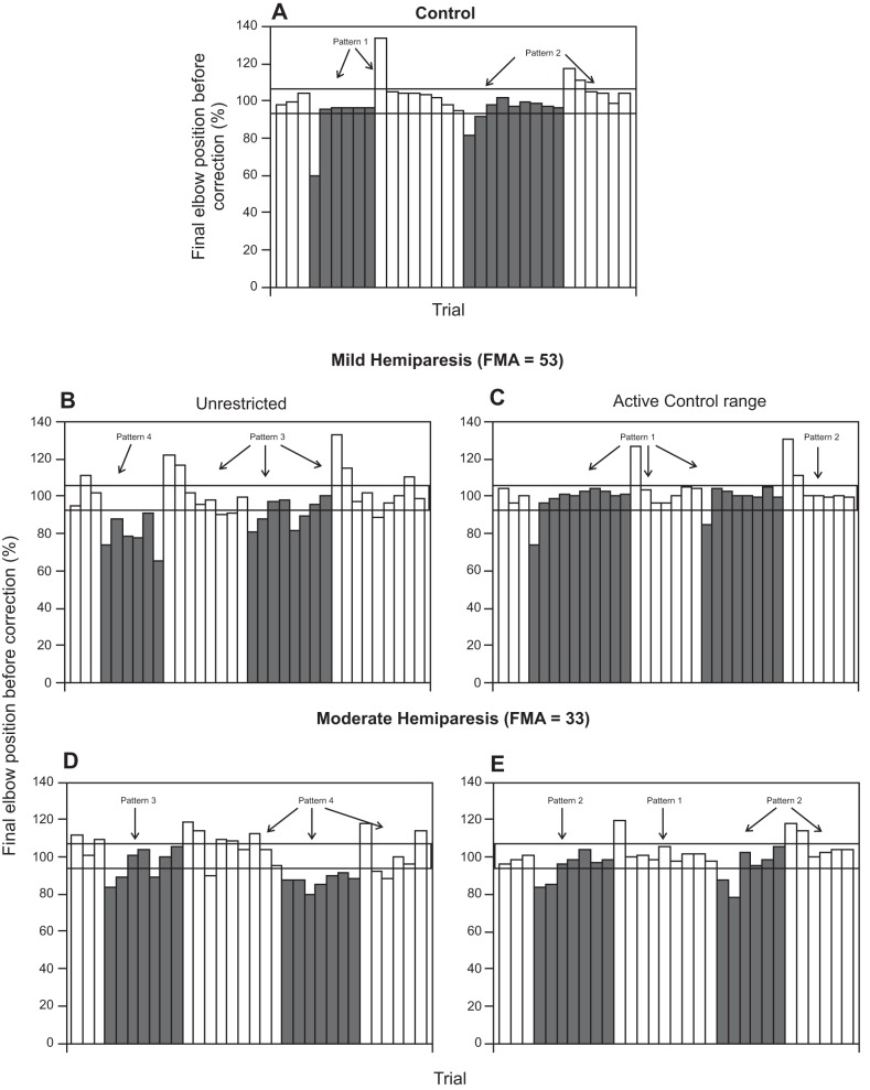
Fig. 8.
Sequence of error correction patterns. Final normalized positions of the primary movement before correction are shown for 4 blocks of trials in a control subject (A) and in subjects with mild (B, C) and moderate (D, E) upper limb hemiparesis when the movements ended in the spasticity zone (B, D) or the active control range (C, E). Open and shaded bars represent trials without or with an opposing load, respectively. The error correction patterns are identified with arrows.

