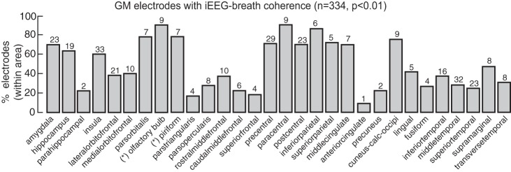
Fig. 4.
Percentage of gray matter electrodes within each brain area showing iEEG-breath coherence above the threshold (99th percentile). Numbers above each bar represent the total number of electrodes within that area showing the effect. Asterisks indicate electrode locations after correction using FreeSurfer parcellation (see materials and methods). Cuneus-calc-occipi, cuneus-calcarine-occipital.

