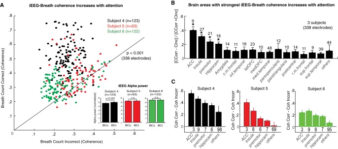
Fig. 9.
Attention to breathing increases iEEG-breath coherence in brain areas related to interoception. A: iEEG-breath coherence values at the respiratory rate during correctly (y-axis) and incorrectly (x-axis) reported breath-counting periods across the population of gray matter electrodes (n = 338 in 3 subjects). Values were increased during correct periods in the 3 subjects collectively (P < 0.001, Wilcoxon’s signed-rank test) and in each subject separately. Black line represents unity. Inset: normalized iEEG alpha power for the 2 conditions and 3 subjects averaged across electrodes sites. None of the subjects showed significant differences between correct and incorrect reports, although there was a trend in subject 4 (P = 0.055) with slightly larger alpha power during incorrect reports. B: distribution of the effect across brain areas. Graph shows mean normalized differences in coherence between correctly and incorrectly reported counting periods. To normalize coherence values across subjects, the difference between correct and incorrect blocks for each subject was divided by their sum. Numbers above bars represent the total number of electrodes within the indicated area. C: coherence values in each subject and brain area (coherence correct – coherence incorrect). The strongest effect was observed in the anterior cingulate cortex, insula, premotor cortex, and hippocampus, and this pattern was consistent across subjects. Other brain areas showed smaller effects. *P < 0.05. Acc, anterior cingulate cortex. Bars indicate means, and error bars indicate SE.

