Fig. 2.
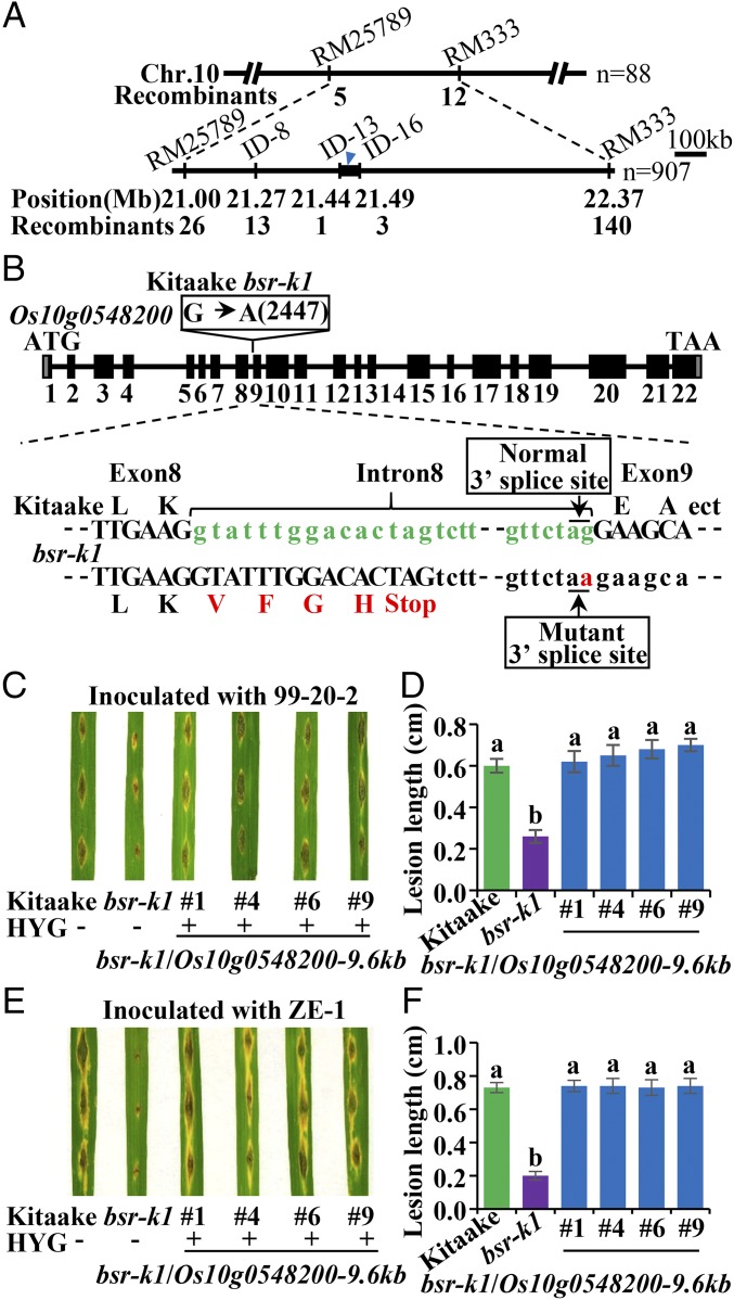
Positional cloning of bsr-k1. (A) Fine mapping of the bsr-k1 locus. The molecular markers and the number of recombinants are indicated. (B) Structure of the Bsr-k1 gene and the mutation in bsr-k1. Filled boxes indicate exons (numbered 1–22) of Os10g0548200. The change from G to A in Os10g0548200 in bsr-k1 is indicated in the open box above exon 9. Nucleotide sequences between exon 8 and exon 9 in Kitaake and bsr-k1 with deduced amino acid sequences are shown below. The substitution in bsr-k1 (in red) abolishes the 3′ splice site of intron 8, resulting in premature termination of translation. (C–F) Blast-resistance test of Bsr-k1–complemented lines. (C and E) Photographs of four independent representative lines at 7 dpi with blast fungal isolates 99-20-2 (C) and ZE-1 (E). (D and F) Lesion lengths (mean ± SD, n > 10) of the complemented lines, bsr-k1, and Kitaake inoculated with blast isolates 99-20-2 (D) and ZE-1 (F). Different letters above bars indicate significant differences (P < 0.05, Tukey’s test). This punch inoculation was repeated twice with similar results.

