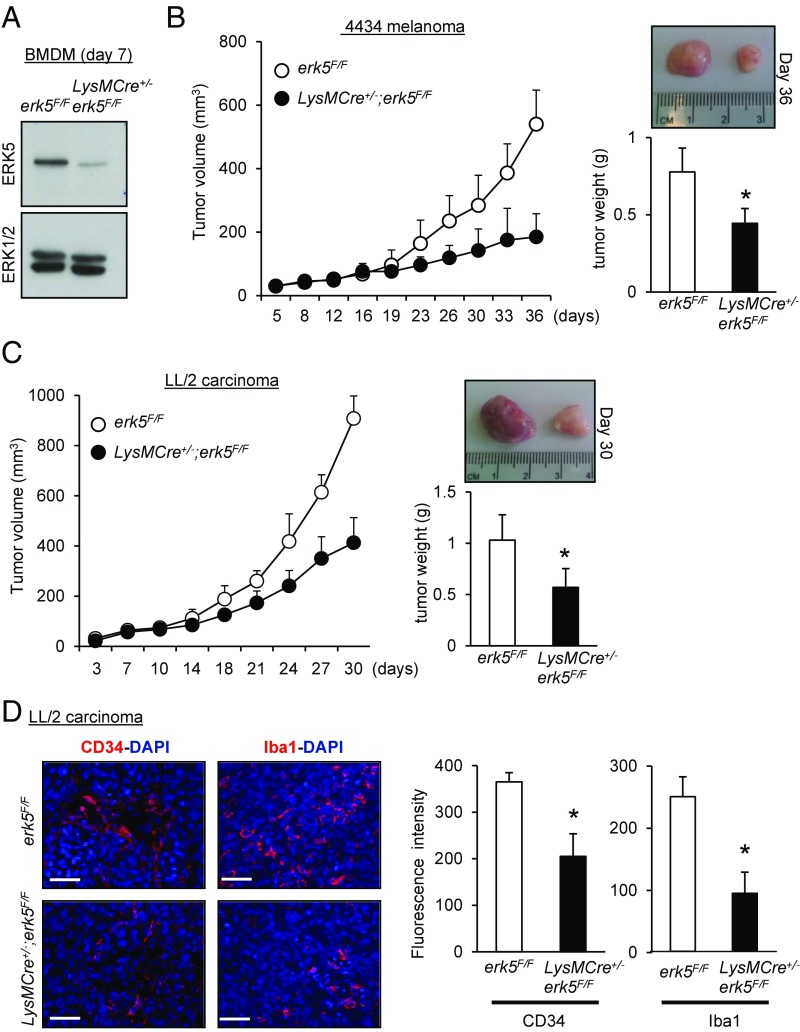
Fig. 2.
ERK5 expression in myeloid cells supports tumor growth in vivo. (A) Immunoblot analysis comparing the level of ERK5 expressed in macrophages obtained from the bone marrow of erk5F/F and LysMCre+/−;erk5F/F animals. Total ERK1/2 expression was used as a loading control. (B and C) The 4434 melanoma (B) or LL/2 carcinoma (C) cells were s.c. implanted into the back of erk5F/F mice that did or did not carry the LysMCre transgene. The growth of tumor grafts was monitored over time after initial cell injection. The data correspond to the mean of tumor volume ± SD (n = 5 mice in each group). Representative pictures of tumor grafts and the mean of tumor weight ± SD excised from erk5F/F and LysMCre+/−;erk5F/F mice killed at the end of the experiment are shown. (D) Sections of carcinoma grafts were analyzed by immunofluorescence using specific antibodies to CD34 (Left) and the pan macrophage marker Iba1 (Right). The immune complexes were detected with a secondary antibody conjugated to Cy3 (red). DNA was stained with DAPI (blue). (Scale bars, 50 μm.) The immunofluorescent signal corresponding to CD34 or Iba1 was quantitated with ImageJ. The data correspond to the mean ± SD (n = 3 tumors). *P < 0.05 (compares tumor grafts from erk5F/F and LysMCre+/−;erk5F/F mice).

