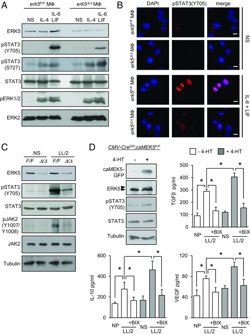
Fig. 6.
STAT3 phosphorylation at Tyr705 in macrophages is dependent on ERK5 signaling. (A–C) After starvation in 1% FBS medium overnight, erk5F/F and erk5Δ/Δ macrophages were stimulated with IL-6 and LIF, or IL-4, or exposed to LL/2-conditioned medium (LL/2) for 30 min, as indicated. Nonstimulated (NS) macrophages were used as controls. (A and C) Protein lysates were analyzed by immunoblot with indicated antibodies. Similar results were obtained in two independent experiments. (B) Immunofluorescence was performed with a specific antibody to pSTAT3(Y705). The immune complexes were detected with a secondary antibody conjugated to Cy3 (red). DNA was stained with DAPI (blue). (Scale bars, 10 μm.) (D) CMV-CreER;caMEK5F/F macrophages were mock-treated (−) or incubated with 4-HT (0.1 μM; +) at day 5 in culture for 48 h. Protein lysates were analyzed by immunoblot with antibodies against GFP, ERK5, pSTAT3(Y705), STAT3, and tubulin. Similar results were obtained in two independent experiments. Alternatively, the cells were incubated with BIX02189 (1 µM; BIX), as indicated, for 2 h before being exposed to LL/2-conditioned medium (LL/2). Nonpolarized (NP) macrophages were used as controls. After 36 h, macrophages were rinsed with PBS and cultured in DMEM supplemented with 5% FBS for a further 24 h. The amount of TGFβ, IL-10, and VEGF secreted in supernatants was quantified by ELISA. The data represent the mean ± SD of two independent experiments. *P < 0.01 (compares differences between treatments).

