Fig. 7.
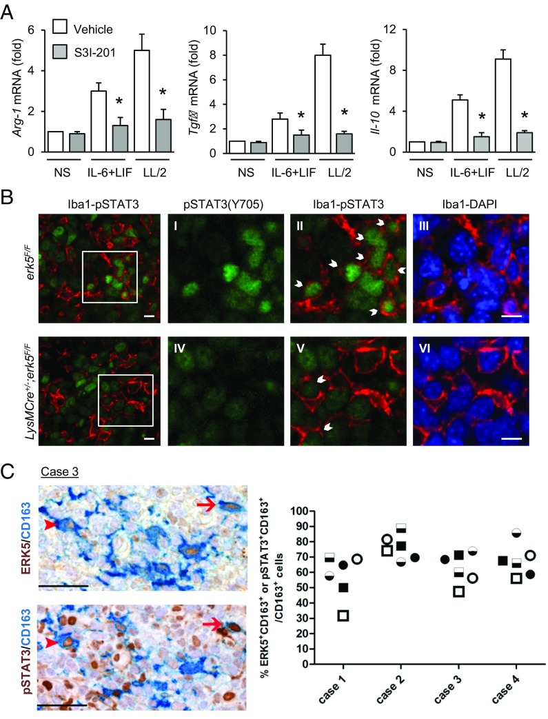
STAT3 is phosphorylated at Tyr705 in TAMs. (A) erk5F/F macrophages were preincubated with S3I-201 (50 μM; Selleck) for 2 h before being stimulated with IL-6 and LIF or with LL/2-conditioned medium (LL/2) for 1 h. Nonstimulated (NS) macrophages were used as controls. Expression of M2 markers was analyzed by qPCR. The data correspond to the mean ± SD of three independent experiments performed in triplicate. *P < 0.05 [compares cells mock-treated (vehicle) vs. treated with S3I-201]. (B) Carcinoma grafts excised from erk5F/F and LysMCre+/−;erk5F/F mice were processed by immunofluorescence with specific antibodies to pSTAT3(Y705) or to the pan macrophage marker Iba1. The immune complexes were detected with Fluorescein Avidin D (green) or Texas Red Avidin D (red). DNA was stained with DAPI (blue). (Scale bars, 10 μm.) B, I–VI are digital magnifications from the corresponding microphotograph. (Scale bars, 15 μm.) Arrows indicate pSTAT3(Y705) positive macrophages. (C) Serial sections obtained from biopsies of human SCC (four cases) were immunostained for ERK5 (brown; Upper) or pSTAT3(Y705) (brown; Lower), coupled with the macrophage marker CD163 (blue). Two HPFs are shown, and the arrows/arrowheads identify two ERK5+pSTAT3+CD163+ triple-positive cells. (Original magnification: 400×; scale bars, 50 μm.) The frequency of double-positive macrophages for ERK5 (circle) or pSTAT3 (square) in three distinct corresponding tumor areas (indicated by black, black/white, and white) is reported in the graph.

