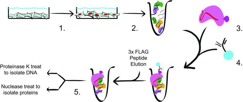
Fig. 1.
Layout of the CLASP. Graphic depiction of the CLASP method. (1) Cells of interest are crosslinked with a crosslinker of choice. (2) Small fragments of chromatin are generated by mechanical shearing of the fixed cells. (3) Recombinant dCas9-3×FLAG loaded with the chosen guide RNA is added to the chromatin mixture. (4) Anti-FLAG antibody conjugated to resin is added to RNP/chromatin and washed; enriched chromatin is eluted with 3×FLAG peptide. (5) Through the use of either Proteinase K or nuclease treatment, enriched DNA or protein samples can be isolated for downstream applications.

