Fig. 1.
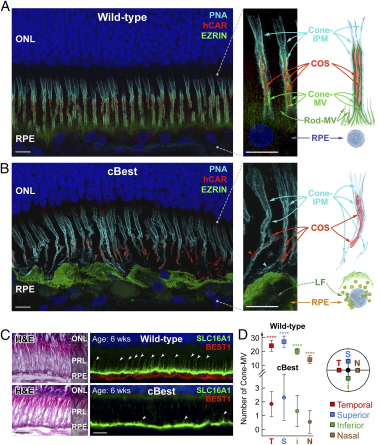
Retina-wide pathology of RPE apical microvillar projections associated with BEST1 mutations. (A and B) Confocal images illustrating the molecular pathology of cBest (R25*/R25*; 89 wk) (B) compared with wild-type (A) (42 wk). Retinal cryosections immunolabeled with anti-EZRIN (green) and human cone arrestin (red) and combined with peanut agglutinin lectin (cyan) and DAPI (blue) detail the structural alterations underlying loss of the native extracellular compartmentalization of cone photoreceptor outer segments (B). The RPE apical marker, EZRIN, recognizes both the smaller hair-like projections capping rod OS tips (rod-MV) as well as the more complex and substantial cone-associated MV (cone-MV). The bilayered extracellular ensheathment associated with a single cone OS (COS) is shown in the magnified photomicrographs (Right), along with the corresponding schematic representation. LF, lipofuscin. (Scale bars, 10 μm.) (C) Representative photomicrographs of 6-wk-old canine wild-type and cBest-mutant (R25*/P463fs) retinas immunolabeled with anti-BEST1 (red) and anti-SLC16A1 (green); white arrowheads point to a subset of cone-MV. H&E, hematoxylin & eosin staining; PRL, photoreceptor IS/OS layer. (Scale bars, 20 μm.) (D) Quantification of cone-MV numbers across the retina between cBest-mutant and age-matched control eyes. The y axis represents the average number of cone-MV per square millimeter for each color-coded retinal region examined (i, inferior; N, nasal; S, superior; T, temporal) with error bars ± SD (****P < 0.0001; n = 80 regions of interest per eye).

