Fig. 1.
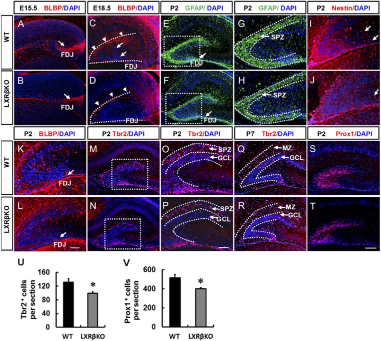
Defective subpial neurogenic zone, abnormal radial glial scaffolding, and decreased granule cell production in LXRβ-null mice. At E15.5 (A and B), BLBP + RGCs formed a new neurogenic zone in the subpial region of the FDJ. Loss of LXRβ decreased BLBP-positive cells in the FDJ. Arrows in A and B indicate the FDJ. At E18.5 (C and D), BLBP+ RGCs crossed the hilus and formed the hippocampal fissure, and loss of LXRβ decreased BLBP-positive cells in the hippocampal fissure. Arrowheads in C and D indicate hippocampal fissure; arrows in C indicate transhilar radial glial scaffold. At P2, GFAP+ processes were more enriched in the transient SPZ of WT animals (E and G) compared with the mutants (F and H). Arrows in E and F indicate FDJ. The SGZ started to take shape. BLBP-positive (K and L) and Nestin-positive (I and J) cells began to seed the nascent SGZ. Loss of LXRβ decreased BLBP-positive and Nestin-positive cells. Arrows in I and J indicate the transhilar radial glial scaffold, arrows in K and L indicate the FDJ. Tbr2+ cells were mainly localized in the SPZ of control animals (M and O), which was decreased in the mutant DGs (N and P) at P2. By P7, as most of the Tbr2+ cells were localized in the GCL, fewer cells were observed in the GCL in the mutant DGs (R) compared with WT controls (Q). At P2, Prox1+ granule cells were decreased in the DG of mutants (T) compared with WT controls (S). Quantitative analysis of the number of Tbr2+ (U) and Prox1+ (V) cells in the DG (n = 3, *P < 0.05, Student’s t test) (G, H, O, and P). Images are higher-power views of the boxed areas in E, F, M, and N. Data are presented as mean ± SEM. (Scale bars: T for A–F, M, N, and Q–T, 100 μm; L for G–L, O, and P, 50 μm.)

