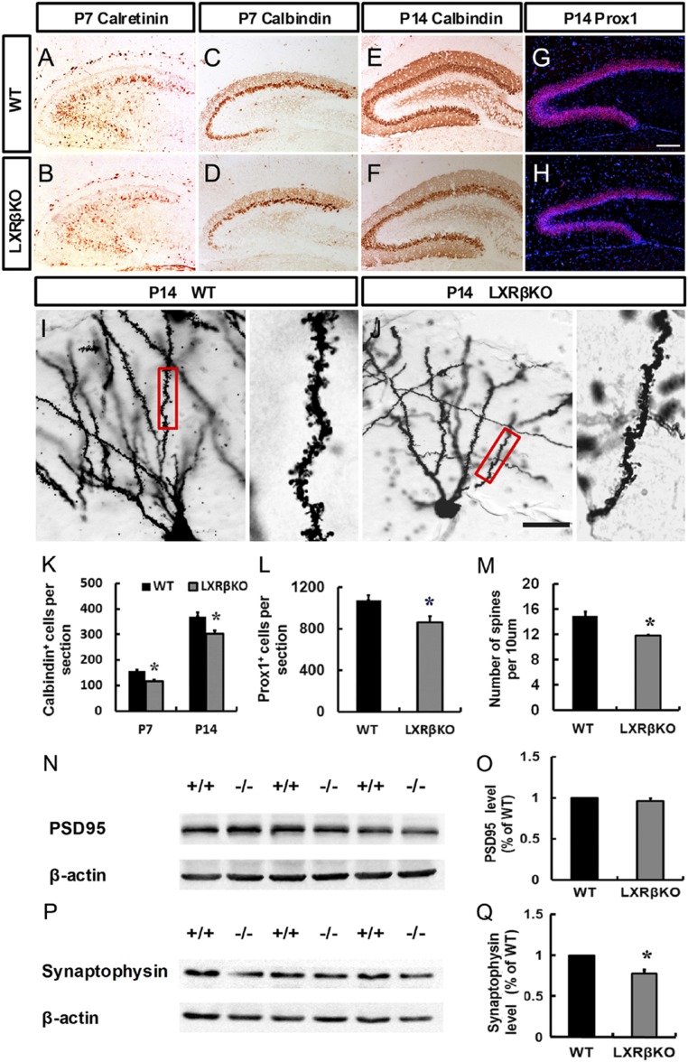
Fig. 4.
Loss of LXRβ disrupted dentate granule cell differentiation. (A and B) Immunohistological analysis of calretinin expression in the DG of control (A) and mutant animals (B) at P7. (C–F) Immunohistological analysis of calbindin expression in the DG of control (C and E) and mutant animals (D and F) at P7 (C and D) and P14 (E and F). (G and H) Immunohistological analysis of Prox1 expression in the DG of control (G) and mutant animals (H) at P14. (Scale bar: G for A–H, 100 μm; J for I and J, 25 μm.) (I and J) Golgi stained dendritic impregnated control and LXRβ mutant hippocampi. Statistical analysis of calbindin-positive (K) and Prox1-positive (L) cells. (M) Statistical analysis of dendritic spine numbers of control and LXRβ mutants at P14. Western blot analysis showed a marked decrease in Synaptophysin (P and Q) in the hippocampal lysates from LXRβKO mice compared with WT at P14. In contrast, no alteration in PSD95 (N and O) level in the hippocampus was detected between WT and LXRβ KO mice. Data are presented as mean ± SEM, n = 3, *P < 0.05, Student’s t test.

