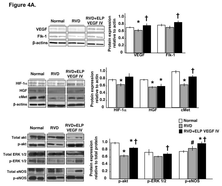
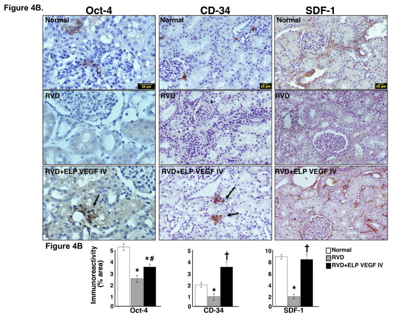
Figure 4. Systemic administration of ELP-VEGF improved angiogenic signaling and stimulated progenitor cells in the stenotic kidney.
4A) Representative renal protein expression (2 bands per group) and quantification of vascular endothelial growth factor (VEGF) and its receptor Flk-1 (top), hypoxia induced factor (HIF)-1α, hepatocyte growth factor (HGF), and HGF receptor cMet (middle), and total and phosphorylated (p)-akt, ERK ½, and endothelial nitric oxide synthase (eNOS, bottom) in normal, RVD and RVD+ELP-VEGF stenotic kidneys. 4B) Representative pictures of immunoreactivity against Oct-4 (40x), stromal-derived factor (SDF)-1 (20x), and CD-34 (20x) and quantification in normal, RVD and RVD+ELP-VEGF stenotic kidneys. * p<0.05 vs. Normal; † p<0.05 vs. RVD; #p>0.1 vs. RVD.


