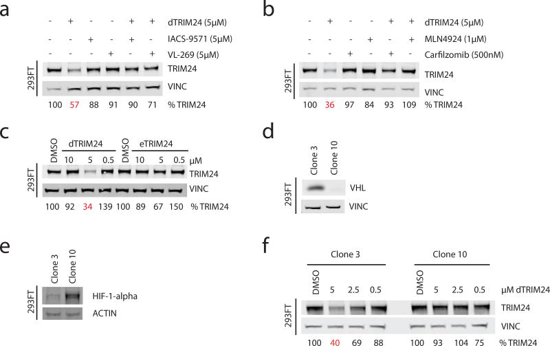
Figure 2. Characterization of the cellular mechanism of degradation of dTRIM24.
(a) Immunoblot of TRIM24 and Vinculin following chemical competition with 5 µM of ligands IACS-9571 and VL-269 with co-treatment of 5 µM dTRIM24 for 24 hours in 293FT cells. (b) Immunoblot of TRIM24 and Vinculin following incubation with carfilzomib (500nM) or MLN4924 (1 µM) with co-treatment of 5 µM dTRIM24 for 24 hours in 293FT cells. (c) Immunoblot of TRIM24 and Vinculin following treatment with the indicated concentrations of dTRIM24 or eTRIM24 for 24 hours in 293FT cells. (d) Immunoblot of VHL and Vinculin in Clone 3 and Clone 10, expanded from single 293FT cells after transient transfection with a guide targeting the VHL locus for CRISPR/Cas9-mediated VHL knockout (antibody: Cell Signaling 68547). (e) Immunoblot of HIF-1α and Actin in Clone 3 and Clone 10. (f) Immunoblot of TRIM24 and Vinculin following treatment of Clone 3 or Clone 10 for 24 hours with the indicated concentrations of dTRIM24. For a–f, percentages were calculated by normalization of the band intensity to the loading control and relative to DMSO. n=3 independently conducted experiments with one representative experiment shown. Full immunoblots shown in Supplementary Fig. 9 and Supplementary Fig. 10.

