
Figure 1.
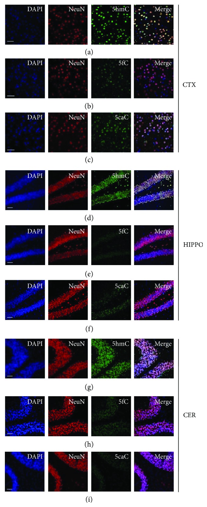
The representative immunofluorescence images of 5hmC, 5fC, and 5caC in the cortex, hippocampus, and cerebellum. Immunofluorescence of 5hmC (a), 5fC (b), and 5caC (c) in the 8 w cortex. Immunofluorescence of 5hmC (d), 5fC (e), and 5caC (f) in the 8 w hippocampus. Immunofluorescence of 5hmC (g), 5fC (h), and 5caC (i) in the 8 w cerebellum. Scale bar, 200 μm.