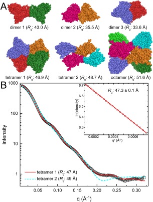
Figure 4.

Case 2: Identifying the correct oligomer from several models. (A) Assembles of the UDP‐galactopyranose mutase from Aspergillus fumigatus derived from PDBePISA analysis of crystal packing (PDB ID 3UTE). Protomers are colored differently for clarity. (B) Experimental SAXS data (open circles) collected at a single protein concentration. The inset shows the Guinier plot. Theoretical curves calculated from tetramer 1 (red solid line; FoXS χ: 3.5) or tetramer 2 (cyan dashed line; FoXS χ: 21) are shown.
