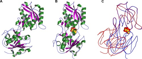
Figure 1.

Overall structure and conformational changes in TpMglB‐2WA. (A) The structure of apo‐TpMglB‐2WA. A ribbons‐style representation of the protein is shown, with helices colored green, β‐strands purple, and regions without regular secondary structure light blue. A large cleft exists between the protein's two domains. (B) The holo‐TpMglB‐2WA structure. Atoms of the bound d‐glucose are shown as spheres, with oxygen atoms in red and carbon atoms in gold. (C) Superposition of the two structures. The C‐lobe was used for alignment purposes. The apo‐structure is shown in red, while that of the holo‐structure is in blue. Simplified traces through the mean Cα positions are shown for each respective structure. The position of d‐glucose in the holo‐TpMglB‐2WA structure is shown for reference.
