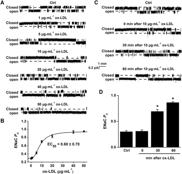
Figure 3.

ox‐LDL stimulates ENaC activity in a dose‐ and time‐dependent manner. (A) Representative ENaC single‐channel currents recorded in endothelial cells from a split‐open aorta either under control (Ctrl) conditions or treated with ox‐LDL at concentrations of 1, 5, 10, 20, 40 and 50 μg·mL−1 for 30 min. (B) ENaC P O were plotted as a function of each corresponding concentration of ox‐LDL and fitted with Pharmacology Standard Curves Analysis using SigmaPlot (n = 6 for each data point). (C) Representative ENaC single‐channel currents recorded in endothelial cells from a split‐open aorta either under control conditions or treated with 10 μg·mL−1 ox‐LDL for 0, 30 or 60 min respectively. (D) Summarized ENaC P O under the conditions shown in (C) respectively (*P < 0.05 vs. respective Ctrl; n = 6).
