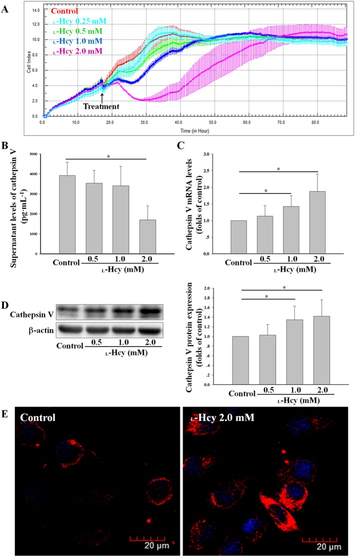
Figure 2.

l‐Hcy promoted the expression of cathepsin V in HUVECs. (A) The effects of 0.25–2.0 mM l‐Hcy on HUVECs viability/proliferation were assessed by RTCA. (B) HUVECs were incubated with 0.5–2.0 mM l‐Hcy for 24 h; the supernatant levels of cathepsin V were determined by ELISA. (C–E) The expression of cathepsin V in HUVECs described in (B) was assessed by real‐time PCR, Western blot and confocal microscopy. The data shown are the mean ± SD of five independent experiments. * P < 0.05.
