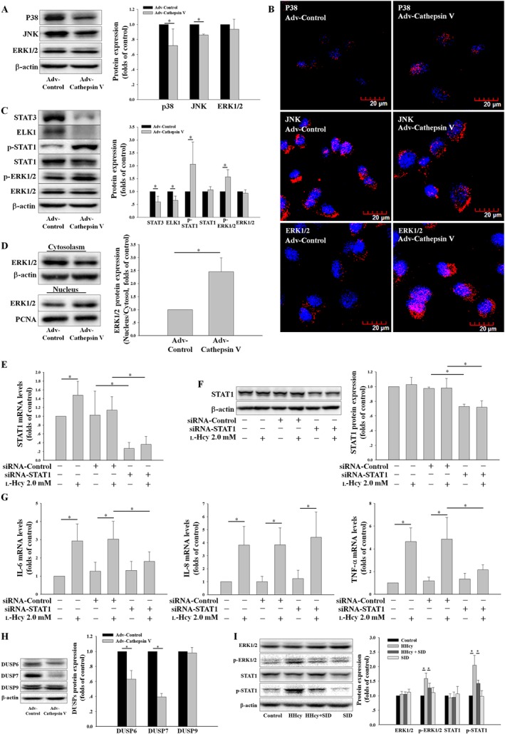
Figure 6.

The ERK1/2/STAT1 pathway is involved in cathepsin V‐induced inflammatory cytokine expression. (A, B) The protein expression and localization of ERK1/2, p38 and JNK induced by Adv‐Cathepsin V were assessed by Western blot and confocal microscopy. (C) The protein expression of STAT3, ELK1, STAT1, phosphor‐STAT1 (p‐STAT1), ERK1/2 and phosphor‐ERK1/2 (p‐ERK1/2) were determined by Western blot. (D) The nuclear translocation of ERK1/2 was detected by Western blot. (E, F) HUVECs were transfected with control (siRNA‐Control) or STAT1 siRNA (siRNA‐STAT1) for 24 h and then co‐incubated with 2.0 mM l‐Hcy for 24 h, the STAT1 expression was assessed by real‐time PCR and Western blot. (G) HUVECs described in (E) were used to assay the mRNA levels of IL‐6, IL‐8 and TNF‐α by real‐time PCR. (H) The protein expression of DUSP6, DUSP7 and DUSP9 induced by Adv‐Cathepsin V were assessed by Western blot. (I) Control mice and hyperhomocysteinaemic mice were separately treated with SID or vehicle, the protein expression of ERK1/2, p‐ERK1/2, STAT1 and p‐STAT1 in the thoracic arteries were assessed by Western blot. The data shown are the mean ± SD of five independent experiments or five animals per treatment. * P < 0.05.
