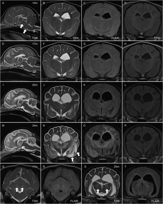
Figure 1.

MR images at 14 months (m) (A–D), 17 months (E‐H, Q, and R), 25 months (I–L, S, and T), and 34 months of age (M–P). From left in each row (A–P), mid sagittal T2‐weighted (T2W) image, transverse images of T2W, fluid attenuated inversion recovery (FLAIR), and T1‐weighted at the level of thalamus. In the bottom row, transverse T2W and FLAIR at the level of cerebellum (Q and R) and same sequences at the level of mid brain (S and T). At 14 m, diffused hyperintense area comparing to cerebral cortex are observed in the brain stem on mid‐sagittal T2W (a; arrows) and cerebral white matter (WM) on transverse T2W (B) and FLAIR (C). At 17 m, hyperintense area in the cerebral WM are obvious on T2W and FLAIR images (F and G). Bilateral hyperintense lesion on T2W and FLAIR were observed in the dentate nucleus (Q and R; arrows). Cerebral and cerebellar atrophy are progressed at 25 m (I, J, K, L). Bilateral hyperintense lesion are observed in the medial geniculate nucleus (bodies) on T2W and FLAIR (S and T; arrows). T2W and FLAIR hyperintense lesion in cerebral WM and thalamus are prominent (M, N, O). Some part of cerebral WM in left temporal lobe (piriform lobe) showed hyperintensity on T2W, and hypointensity on T1W and FLAIR (N, O, P; arrows).
