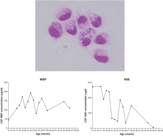
Figure 3.

Analysis of cerebrospinal fluid (CSF). Some of mononuclear cells in CSF contained eosinophilic granules within the cytoplasm. These cells could be seen in CSF until 26 months of age. Follow‐up measurement of neuron specific enolase and myelin basic protein revealed increased concentration with time associated with neurons and myelin destruction.
