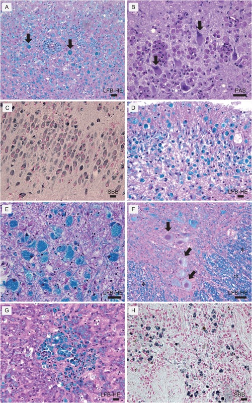
Figure 4.

Histopathology of dog with SD. (A) Cerebral cortex stained with Luxol fast blue and, hematoxylin and eosin (LFB‐HE). There is severe neuron loss and remaining neurons are swollen with LFB positive granular inclusions (arrows). Microglia also contained LFB‐positive materials. (B) Cerebral cortex stained with periodic acid‐Schiff (PAS). Inclusions within cytoplasm were PAS‐positive (arrows). (C) Hippocampus stained with Sudan black B (SBB) revealed inter‐cellar inclusions are SBB‐positive. (D) Cerebellum, LFB‐HE. (E) Brain stem, LFB‐HE. Though neurons are preserved they are severely swollen with LFB positive granular inclusions. (F) Spinal cord: LFB‐HE. Well‐preserved myelin in white matter (*) and severely swollen neurons with LFB positive inclusions were observed (arrows). (G) Liver, LFB‐HE. LFB‐positive cells in the liver. (H) Spleen, SBB‐positive cells. Scale Bar represents 100 μm for all panels.
