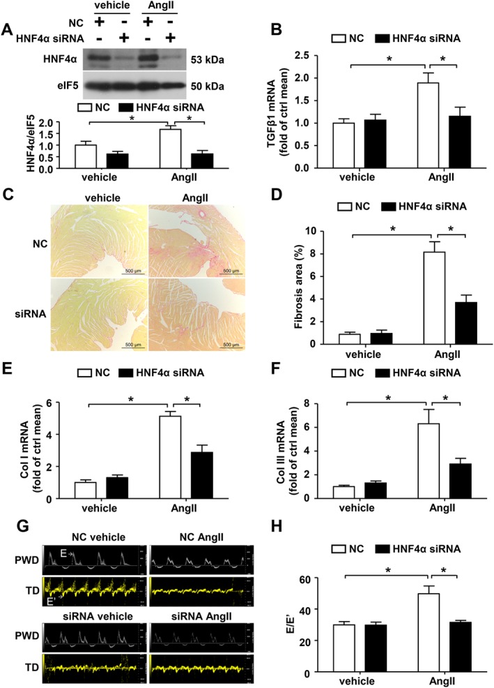
Figure 3.

HNF4α mediates AngII‐induced TGFβ1 expression and cardiac fibrosis in vivo. The mice were treated with negative control (NC) or HNF4α siRNA followed by AngII infusion. (A) Western blot analysis of HNF4α expression in the hearts. (B) Quantitative real‐time PCR analysis of TGFβ1 mRNA expression in heart lysates. (C) Representative micrographs of Sirius red‐stained heart sections; the red area represents collagen. Bars = 500 μm. (D) Quantification of the fibrotic area is expressed as a percentage of the total cardiac area. Collagen I (E) and collagen III (F) mRNA expression were measured via real‐time PCR analysis. (G) Representative pulsed wave Doppler (PWD) images across the mitral flow and tissue Doppler (TD) images of the mitral valve ring on the seventh day of AngII infusion in wild type mice. (H) E/E’: Bars represent means ± SEM of six mice per group. *P < 0.05. Two‐way ANOVA with the Bonferroni post hoc test was used (A, B and E). Welch's ANOVA with post hoc Games–Howell test was used in (D, F and H).
