Figure 1.

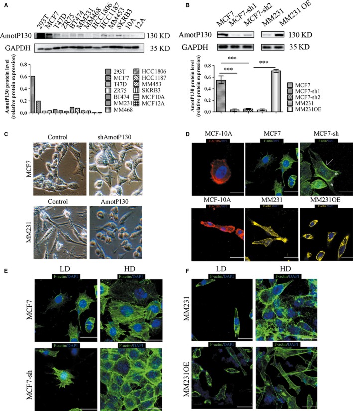
AmotP130 was differential expressed in breast cell lines and correlated with cell morphology and cytoskeleton arrangement. MCF7‐sh, the AmotP130 silenced MCF7 cells; MM231, MDA‐MB‐231 cells; MM231 OE, the AmotP130 overexpressed MDA‐MB‐231 cells; LD, low cell density; HD, high cell density. A, Western blot analysis of AmotP130 expression in positive control cell (293T) and breast cancer cell lines. B, Western blot verification of knockdown and overexpression efficiency of AmotP130 in MCF‐7 and MM231 cells. C, Images of different cell morphology depending on the AmotP130 expression status under 200× field. D, Representative fluorescent images of F‐actin cytoskeleton labelled by phalloidin showed different actin microfilaments arrangement depended on the AmotP130 expression status. E, Representative fluorescent images of F‐actin cytoskeleton labelled by phalloidin in MCF7 and MCF7‐sh cells under LD and HD. F, Representative fluorescent images of F‐actin cytoskeleton labelled by phalloidin in MM231 and MM231OE cells under LD and HD. Scale bar 10 μm. Values were mean ± SD. ***P < .001
