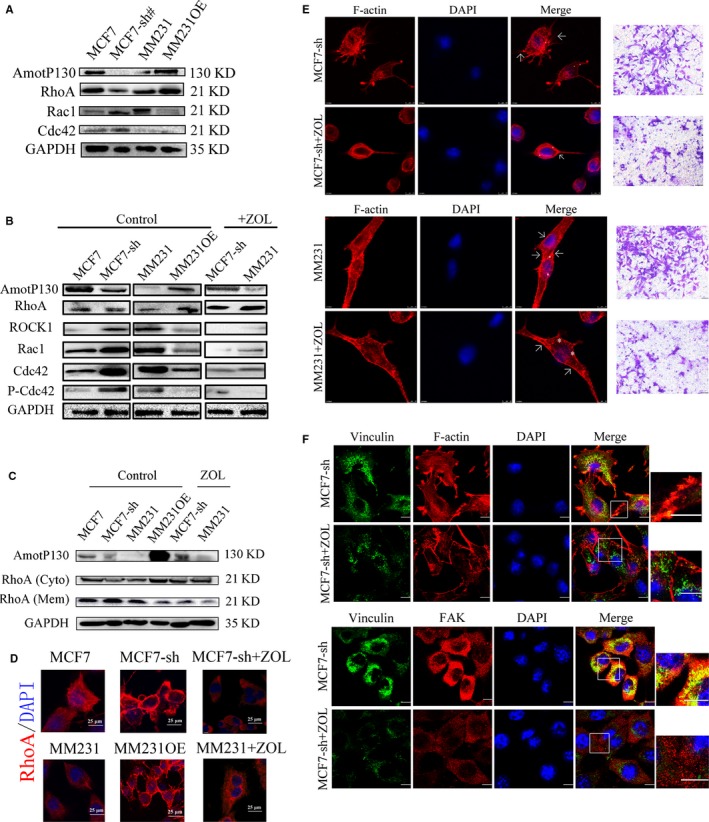
Figure 5.

Rho signalling pathway inhibition suppressed actin filaments polymerization and reduced the invasion ability induced by AmotP130 down‐regulation. MCF7‐sh, the AmotP130 silenced MCF7 cells; MM231 OE, the AmotP130 overexpressed MDA‐MB‐231 cells, ZOL, zoledronic acid. A, Western blot analysis of total protein expression of RhoA, Cdc42, Rac1 in MCF7, MCF7‐sh, MM231 and MM231OE cells. B, Western blot analysis of RhoA, Rock1, Cdc42, Rac1, P‐Cdc42/Rac1 total protein expression in cell lines as above. After ZOL 70 μmol L−1 treatment for 24 h, these protein were tested again in MCF7‐sh and MM231 cells. C, RhoA membrane and cytosol protein expression were analysed before and after ZOL 70 μmol L−1 treatment for 24 h. D, Immunofluorescence assays showed the translocation of RhoA in studied cells before and after ZOL 70 μmol L−1 treatment for 24 h. Scale bar 25 μm. E, Immunofluorescence and Transwell migration assays showed the arrangement of F‐actin and invasion ability of MCF7‐sh cells (upper panel) and MM231 cell (down panel) before and after ZOL 70 μmol L−1 treatment for 24 h. The asterisk labelled scattered actin and arrow labelled fibres gathered at cell cortex. Invading cells were stained and photographed under 200× field. F, Immunofluorescence assay showed double staining experiments of FAK and vinculin in MCF7‐sh cells (upper panel), and the organization of vinculin and F‐actin (down panel) before and after ZOL 70 μmol L−1 treatment for 24 h. Scale bar 10 μm
