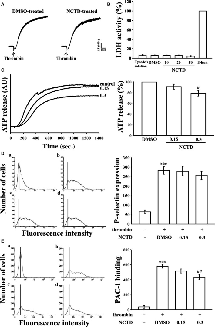
Figure 2.

Effects of NCTD on cytotoxicity, lactate dehydrogenase (LDH) release, ATP‐release reaction, surface P‐selectin expression and integrin αII bβ3 activation in human platelets. (A) Washed platelets were preincubated with the solvent control (0.1% DMSO) or NCTD (5 μM) for 10 min. and subsequently washed two times with Tyrode solution; thrombin (0.01 U/ml) was then added to trigger platelet aggregation. (B) Washed human platelets (3.6 × 10⁸ cells/ml) were preincubated with NCTD (10, 20 and 50 μM) for 20 min., and a 10‐μl aliquot of the supernatant was deposited on a Fuji Dri‐Chem slide LDH‐PIII as described in ‘Methods’. (C) Moreover, washed platelets (3.6 × 10⁸ cells/ml) were preincubated with NCTD (0.15 and 0.3 μM) or the solvent control (0.1% DMSO), and 0.01 U/ml thrombin was then added to stimulate the ATP‐release reaction (AU; arbitrary unit). For other experiments (D‐E), resting platelets (a) or platelets (3.6 × 10⁸ cells/ml) were preincubated with the solvent control (b, 0.1% DMSO) or NCTD (c, 0.15; d, 0.3 μM) and the FITC‐conjugated anti‐P‐selectin mAb (2 μg/ml) or the PAC‐1 mAb (2 μg/ml) for 3 min. and then stimulated by thrombin (0.01 U/ml) for another 5 min. The suspensions were then assayed for fluorescein‐labelled platelets on a flow cytometer (FACScan system, Becton Dickinson). Profiles in (A) are representative of four independent experiments. Data in (B‐E) are presented as means ± standard errors of the means (n = 4). ***P < 0.001, compared with the resting group; # P < 0.05 and ## P < 0.01, compared with the 0.1% DMSO‐treated group.
