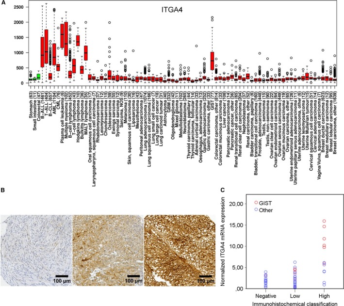
Figure 1.

(A) A box–whisker plot showing the relative ITGA4 mRNA expression in histopathologically normal gastric, small intestine and colorectal tissue (green boxes) and in different types of cancer (red boxes). The number of samples studied is indicated in the brackets. (B) Examples of negative, low and high ITGA4 expression in GIST tissue samples (magnification ×200). (C) Association between ITGA4 protein expression at immunohistochemistry and the normalized ITGA4 mRNA expression (P < 0.001) determined from the same samples with qPCR. Panel A is modified from IST Online™ (ist.medisapiens.com).
