Figure 1.

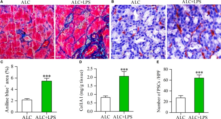
LPS was associated with increased expression of collagen and activation of PSCs. Aniline blue staining showing collagen deposition in the pancreas (blue) of alcohol (ALC, 15 g/kg/d)‐fed rat and ALC rat plus LPS (3 mg/kg once a week for 3 weeks) repeated injections (ALC + LPS) (A); Immunostaining showing the activated PSCs in the pancreas of ALC rat and ALC + LPS rat (B); The area of collagen deposition (C) or the content of Col1A1 (D) or the number of activated PSCs (E) in the pancreas of ALC + LPS rats was significantly more than that of ALC rats. Student's t test, ***P < .001 vs ALC rats (n = 11/group)
