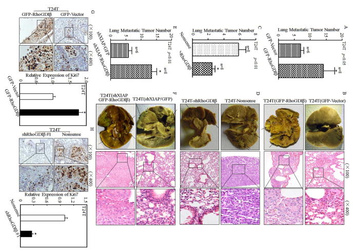
Figure 6. RhoGDIβ was essential for promoting T24T cell lung metastasis.
The T24T/pEGFP, T24T/pEGFP-RhoGDIβ, T24T/nonsense, T24T/shRhoGDIβ, T24T(shXIAP/pEGFP), T24T(shXIAP/pEGFP-RhoGDIβ) cells were intravenously inoculated into nude mice as described in “Methods”. (A, C&E) The lung metastatic tumor numbers of T24T/pEGFP, T24T/pEGFP-RhoGDIβ (A); T24T/nonsense, T24T/shRhoGDIβ (C); and T24T(shXIAP/pEGFP), T24T(shXIAP/pEGFP-RhoGDIβ) (E) were analyzed. (B, D&F) Representative images of the lungs and lung surface metastatic foci as indicated were shown after fixation in a neutral-buffered formalin/Bouin’s fixative solution (left panel), and the histologic appearance of lung metastases were analyzed using H&E staining (right panel), respectively. (G&H) Ki-67 protein expression of lung metastatic tumor tissues was detected using immunohistochemical staining analysis (G&H, left panel) and the quantitative analysis was done indicated paired groups (G&H, right panel). The results were presented as mean ± SD from at least triplicate experiments and asterisk (*) indicated a significant difference (P <0.05).

