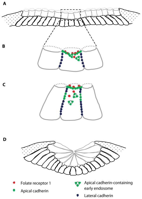
Figure 1.
Model of the role of folate receptor 1 in neural plate folding. (A) Two layers (grey – superficial, black – deep) of Xenopus laevis neural plate cells at the beginning of folding show a cuboidal and non-polarized morphology. Speckled regions represent non-neural ectoderm. (B) In superficial neural plate cells folate receptor 1 (red) and cadherins (green) localize to the apical membrane. Cadherins are also enriched in the cell lateral border (blue). Circles inside the cell represent early endosomes containing apical cadherin. (C) Folate receptor 1-dependent cadherin endocytosis results in redistribution of adhesion molecules and membrane from the cell apex to the lateral cell border, facilitating constriction of the apical pole and elongation of the neural plate cell. (D) Apical constriction along with other morphogenic behaviors such as cell elongation and intercalation lead to folding of the neural plate. Model derived from Balashova et al., 2017.

