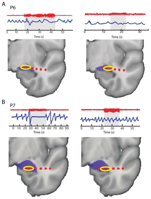
Figure 6.
Amygdala-stimulation-induced apnea is specific to the medial sub-regions of the amygdala. Two patients had electrode coverage spanning the mesial and lateral portions of the amygdala, providing the opportunity to test whether the effect differed across the medial and lateral amygdala sub-regions. In two patients (P6 and P7), we delivered bipolar stimulation to the mesial-most and lateral-most contacts within the amygdala and in both patients, we found that apnea only occurred during stimulation of the medial amygdala. The red traces show the iEEG data from the amygdala, with the blue traces showing the respiratory signal. Below each example is the individual patient’s amygdala electrode locations. The yellow circles indicate which two electrodes were stimulated in each instance.

