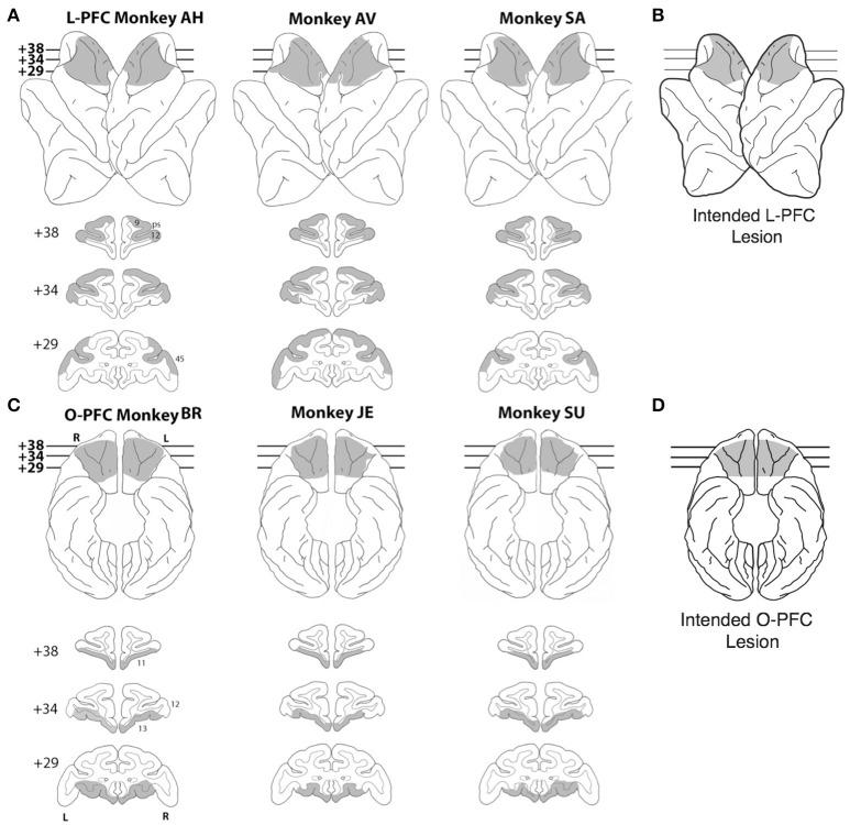
Figure 1.
Lesions. (A) Lateral view (top) and coronal sections (bottom) of a standard rhesus monkey brain showing the extent of the LPFC lesion (shaded regions) in all three monkeys. Areas corresponding to cytoarchitectonic regions 9, 12, and 45 are indicated on coronal sections. (B) Lateral view of the intended LPFC lesion. (C) Ventral view (top) and coronal sections (bottom) of a standard rhesus monkey brain showing the extent of the OFC lesion (shaded regions) in all three monkeys. Areas corresponding to cytoarchitectonic regions 11, 12, and 13 are indicated on coronal sections. (D) Ventral view of the intended OFC lesion. The numerals next to each coronal section in panels (A,C) indicate the distance in millimeters from the interaural plane. ps, principal sulcus; L, left hemisphere; R, right hemisphere.

