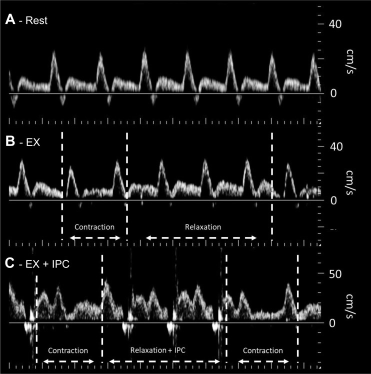
Fig. 4.
Example of the Doppler tracings acquired from the popliteal artery at rest (A), during exercise without compression (B), and during exercise with compression (C). Compared with rest, blood velocity was elevated in both exercise conditions. Muscle contraction during exercise resulted in slight reductions in the Doppler trace, where the application of compression during the relaxation phase is clearly visible as brief periods of reverse velocity followed by an increase in diastolic flow. Note: contrast has been slightly increased to allow for better visualization of changes in the velocity profile.

