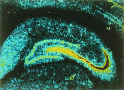
Figure 1.
Distribution of kainate binding sites in the hippocampus. Binding site density is color-coded with high to low densities represented by red-yellow-blue. The autoradiography was carried out with [3H]kainate and shows a high labeling density localized to the stratum lucidum, the termination zone for mossy fibers. [Reprinted with permission from ref. 12 (Copyright 1982, Elsevier Science).]

