Figure 5.
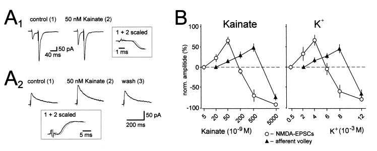
Bidirectional control of synaptic transmission by kainate and presynaptic membrane potential. (A1) Averaged traces of AMPAR EPSCs recorded at −70 mV holding potential in the presence of picrotoxin (100 μM). Kainate (50 nM) increases the amplitude of the first synaptic current, whereas the second is unchanged, thereby decreasing paired pulse facilitation. Note that the increase is not associated with a change in the rising phase of the EPSC. (A2) Averaged traces of NMDAR-EPSCs recorded at +30 mV holding potential in the presence of the AMPAR antagonist GYKI 53655 (20 μM) and the GABAA receptor antagonist picrotoxin (100 μM) are shown. Kainate (50 nM) reversibly increases the amplitude of the synaptic current. Note that the increase is not associated with a change in kinetics of the EPSC. (B) Concentration dependency of the effects of kainate and K+ additions on NMDAR-EPSCs and afferent volley size. Note that 20 nM kainate and 2 mM K+ significantly increase the amplitude of the NMDAR-EPSC, whereas the fiber volley is not affected. Note also that 500 nM kainate and 8 mM K+ cause an enhancement of the afferent volley, whereas synaptic transmission is strongly suppressed. n ≥ 5 for each experiment. [Reprinted with permission from ref. 22 (Copyright 2001, American Association for the Advancement of Science, www.sciencemag.org).]

