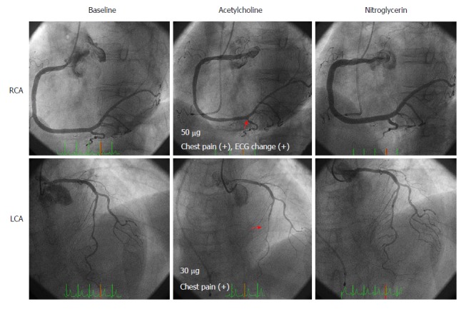
Figure 1.

Coronary angiography and spasm provocation test (SPT) for Case 1. Coronary angiograms show angiographically normal coronary arteries (left upper and lower panels). During the SPT, coronary spasms occurred at the distal segment of the right coronary artery (RCA) at 50 μg of ACh (middle upper panel) and at the mid-segment of the left anterior descending coronary artery (LAD) at 30 μg of ACh (middle lower panel). These coronary spasms resolved after an injection of nitroglycerin (right upper and lower panels). Spastic segments are indicated by arrows.
