Figure 3.
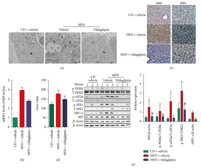
Vildagliptin ameliorated high fat diet induced endoplasmic reticulum (ER) stress. (a) Electron microscope (magnification ×15000) analyses of the ER in livers of mice from CD, HFD, and V-HFD groups. Scale bars represent 2 μm. (b) The mRNA levels of BiP measured by RT-PCR; data were normalized according to β-actin levels. (n = 5 per group). (c) The expression of BiP was assessed using immunohistochemical staining (magnification ×200 and magnification ×400). (d) The semiquantitative analysis of staining intensity was conducted using ImageJ software. (e) ER stress associated markers BiP, p-PERK, p-IRE1α, p-eIF2α, and xBP-1s expression in the livers of mice investigated by western blotting. Each expression level was quantified by densitometry, normalized with β-actin, and the relative phosphorylated protein levels were normalized with the corresponding total protein level. Data was represented as the means ± SD; ∗P < 0.05 relative to CD group; &P < 0.05 relative to HFD group.

