Fig. 3.
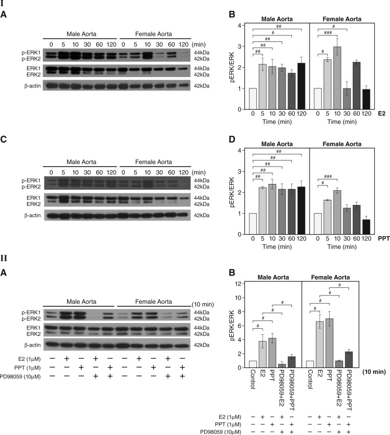
Increased phosphorylation of ERK after 17β-estradiol (E2) or 4,4′,4″-(4-propyl-[1H]pyrazole-1,3,5-triyl)tris-phenol (PPT) treatment in male and female aortas. I,A: representative Western blots for levels of phosphorylated (p)ERK1/2 and total ERK1/2 at different time points after E2 (1 µM) stimulation in aortas. β-Actin is shown as the loading control. I,B: quantitation of I,A. pERK was normalized to total ERK1/2 (n = 3 experiments, #P < 0.05, ##P < 0.01, and ###P < 0.001, E2 vs. vehicle). I,C: representative Western blots for the levels of pERK1/2 and total ERK1/2 at different time points after PPT (1 µM) stimulation in aortas. I,D: quantitation of I,C. pERK1/2 was normalized to total ERK1/2 (n = 3 experiments, #P < 0.05, ##P < 0.01, and ###P < 0.001, PPT treatment vs. vehicle controls). II,A: effect of the ERK inhibitor PD-98059 on pERK1/2 after E2 or PPT treatment in male and female mouse aortas. Representative Western blots are shown for pERK1/2 and total ERK1/2 after E2 (1 µM) or PPT (1 µM) stimulation for 10 min with or without PD-98059 preincubation at 10 µM for 30 min. II,B: effect of the ERK inhibitor PD-98059 on pERK after E2 or PPT treatment in male and female mouse aortas (quantitation of II,A). The level of pERK1/2 was normalized to that of total ERK1/2 (n = 3 experiments, #P < 0.05). Data are expressed as means ± SE.

