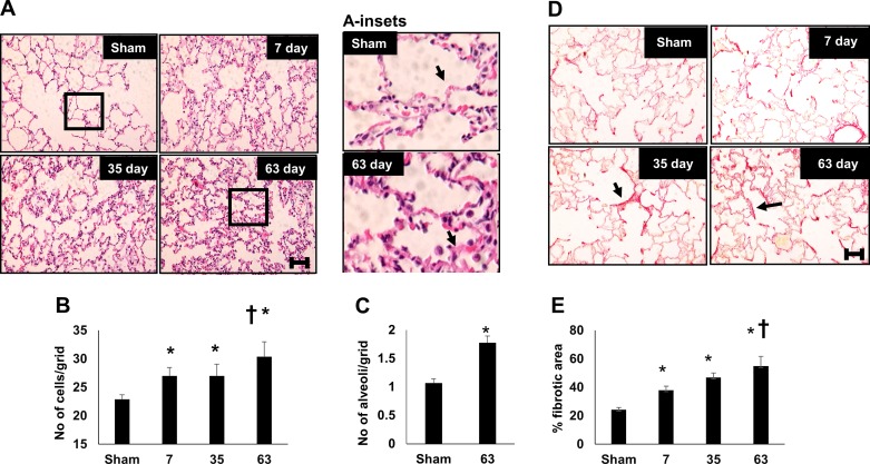
Fig. 5.
Renal ischemia-reperfusion (I/R) injury results in persistent alterations in lung structure. A: representative hematoxylin and eosin (H&E)-stained images of lung parenchyma obtained from rats after sham surgery or after recovery from renal I/R. Inset: a higher magnification is also shown. Quantification of cell density expressed as number per arbitrary grid (B) or the number of alveoli per arbitrary grid (C) is shown. D: representative images of picrosirus red-stained lung cross section. E: percent picrosirius red-stained relative to total cellular area. Magnification bar (100 µm) is shown in bottom panels. Data are means ± SE. For B and E, *P < 0.05 I/R groups vs. sham; †P < 0.05 vs. 7 day post-I/R, by one-way ANOVA and Tukey’s post hoc test. For C, *P < 0.05 by Student’s t-test. (n = 3–4 rats per group).

