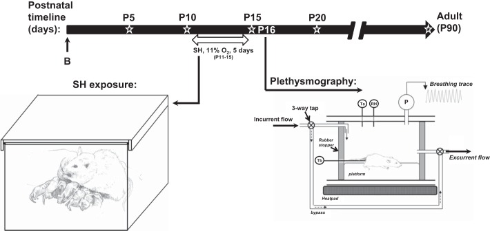
Fig. 1.
Timeline and experimental setup used to expose rats to sustained hypoxia [SH (11% O2, 24 h/day) between postnatal days (P) 11 (P11) and P15]. At the end of P15, rats were removed from the SH chamber, and the acute hypoxic ventilatory response (HVR) was assessed by whole body plethysmography on the following day (P16). Open star designates time points at which semiquantification of Wisteria floribunda agglutinin and aggrecan was carried out within brain stem regions (see Figs. 2 and 3). B, birth.

