Fig. 4.
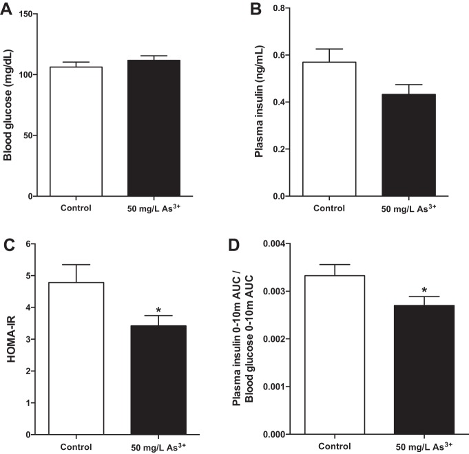
Arsenic exposure induces metabolic dysfunction. A: fasting glucose after a 6-h fast. B: fasting insulin collected from peripheral blood after a 6-h fast. C: homeostatic model assessment of insulin resistance (HOMA-IR) values calculated using fasting blood glucose and fasting plasma insulin measures. D: change in insulin (AUC) relative to the change in blood glucose (AUC) from 0 to 10 min during an IP-GTT; n = 14–16 mice per group (cohort 1). Data are means ± SE; *P < 0.05.

