Fig. 5.
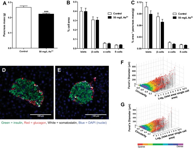
Arsenic exposure does not affect islet morphology or endocrine cell composition. A: pancreas mass measured immediately after each mouse was euthanized. B: histological sections of pancreas from each mouse were immunostained, and endocrine cell area [β-cell, α-cell, δ-cell, and islet (additive)] was calculated relative to total area analyzed. C: islet mass as measured by %cell area multiplied by pancreas mass for each animal. D: representative image of a single islet from control mouse. E: representative image of a single islet from arsenic-exposed mouse. F: scatter plot of individual islet morphology from all control mouse slides. G: scatter plot of individual islet morphology from all arsenic-exposed mouse slides; n = 16 slides per group (cohort 1). Each slide is representative of 1 individual mouse. Data are means ± SE; ***P < 0.001.

